


Skin Analysis
Skin Analysis in Birmingham, Ann Arbor and Detroit
Reveal Skin Imperfections
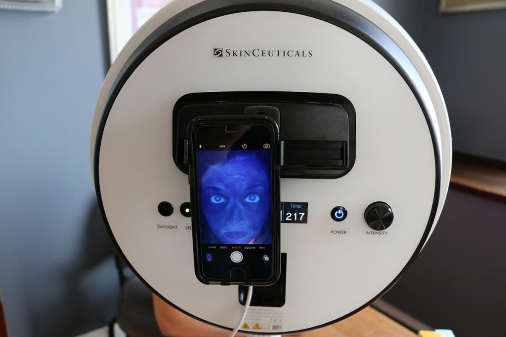
Introducing the next generation of portable, full-face diagnostics: the SkinScope LED
Skinscope LED allows the Boyd team to see visible and underlying skin imperfections including accumulated sun damage (lentigines), oily skin and congested pores, dehydrated and thinner skin areas, uneven texture, and poor desquamation.
What are the Skinscope's Features and Benefits?
• Offers multiple diagnostic options with purer light capabilities by removing the purple ‘haze’ of visible light emitted by wood’s lamp bulbs
• Reveals visible and underlying skin imperfections including accumulated sun damage (lentigines), oily skin and congested pores, dehydrated and thinner skin areas, uneven texture, and poor desquamation
• Enables follow-up engagement by facilitating smartphone photography of diagnostic sessions
• Allows the skincare professional to recommend regimens and products to the client based on diagnostic results
• Helps the skincare professional convey progress to clients after visits and treatments over time
• Incorporates timer settings for each light mode for controlled consultation sessions
• Easy to use, light, and portable
What are the Diagnostic Modes?
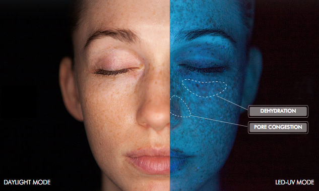
While some skin concerns and imperfections are visible in everyday light, some will only be visible under UV light which highlights damage beneath the skin’s surface by detecting skin’s fluorescence.
The SkinScope LED has two light modes: simulated Daylight mode for reviewing visible skin
conditions and concerns, and a LED-UV light mode for reviewing skin fluorescence (emitted at 320-365nm). Both lights are produced by solid-state UV emitters dispersed by six polished chrome mirrors.
How Does the Skinscope LED Work?
1. DAYLIGHT
The simulated Daylight mode allows for the clear illumination of ‘visible’ concerns to the patient and skincare professional. The diagnostic advisor can pinpoint what concerns the patient has and can highlight areas of redness, irritation, visible dryness, oiliness, wrinkles, and pigmentation.
2. LED-UV 320-365NM
The LED-UV mode illuminates sub-surface imperfections visualized by the fluorescence of the skin. This brings to life concerns that may be faintly visible in daylight but are acutely emphasized under UV light. While healthy skin reflects back UV light creating a blue glow, melanin in the skin absorbs the light showing as dark spots on the surface of the face. Similarly, congested pores give off pink or orange fluorescence, oily skin is visible in a yellow color, and dry flaky skin shines as bright white fluorescence. Large patches of darker blue indicate areas of thinner, dehydrated skin.
What is the Color Guide?
THE SCIENCE OF FLUORESCENCE TECHNOLOGY
Fluorescence is caused when one radiation wavelength is absorbed by a compound which is reflected back at a different wavelength. Certain compounds excite electrons in molecules that change the wavelength energy such that it converts from shortwave UV light to longer wave visible light.
When a very specific range of UV light (320-365 nm) illuminates skin it reacts in different ways based on what it comes in contact with. Melanin absorbs the light showing as an absence of color, but other compounds ‘excite’ follicular fluorescence in the skin, changing the wavelength to colors visible to the human eye. Based on the visible shades that are reflected back from the skin, characteristic diagnoses can be made. Propionibacterium acnes, for example, are a bacterium implicated in acne causation which always glow an orange/pink color. Drier skin flakes from poor desquamation will fluoresce a bright white color. Healthy skin will fluoresce a homogenous light blue, while lipid deficient or thinner skin areas will be indicated by darker shades of blue.
What Else Should Be Considered?
Fitzpatrick skin types 4-6 may be more difficult to assess in the SkinScope LED as a baseline of endogenous melanin exists. You will be able to assess blocked comedones and p-acnes by following the color guide.
•Some deodorants, soaps and lint can fluoresce under LED-UV light.
•Diagnostic assessment can be skewed if the patient is wearing makeup or sunscreen. Ideal conditions call for a clean, makeup and sunscreen-free face and neck.
•Washing the face right before diagnosis can create a false-negative result.
What Diagnostic Support is Used?
•Diagnostic color chart for Daylight and LED-UV with a SkinCeuticals diagnostic step-by-step guide
•SkinCeuticals Diagnostic Worksheet
Pale Blue: Normal and healthy skin
White: Dead skin cells
Dark Blue: Thinner, dehydrated skin
Brown: Pigmentation and dark spots
Yellow: Oily areas of the face*
Dark Pink or Orange: Congested pores and comedones*
Harvard Medical School graduate and double board-certified facial plastic surgeon, Dr. Charles Boyd, and the BOYD team are proud to offer non-surgical cosmetic treatments at BOYD Ann Arbor, Birmingham, Detroit, Michigan. Contact us today to schedule your one-on-one consultation with Dr. Boyd.